A 
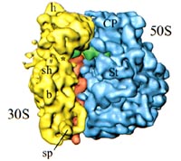
Model of the Ribosome Prepared by Cryo-Electron Microscopy
(Figure 12-13 from Biology by Miller & Levine)

The ribosome is a complex subcellular particle composed of protein and RNA. It is the site of protein synthesis, as described on pages 300-306 of the Dragonfly Book. In recent years, a number of laboratories have advanced the study of the ribosome to the point where we now have detailed atomic level models of the ribosome that explain many of the most important aspects of protein synthesis. One of the leaders of this research is Dr. Joachim Frank of the Wadsworth Institute, who kindly provided the image used in our textbook (shown at left).

In the first few printings of our textbook, this image was described as having been prepared by neutron and X-ray diffraction. That was a mistake. While those are both useful techniques in the study of ribosomes, in fact this image was prepared by Cryo-electron microscopy:

Cryo-Electron Microscopy

Dr. Frank has pioneered a technique known as cryo-electron microscopy, in which biological samples are embedded in a thin layer of ice and then carefully examined in the electron microscope. The technique allows delicate structures like the ribosome to be preserved in their native state, and provides enough resolution (fine detail) to allow scientists to study them closely. In order to do this, thousands of images of individual ribosomes, tilted to a variety of different angles, were combined using an innovative computer program known as SPIDER, also developed by Dr. Frank and his associates. The SPIDER program permits researchers to use the electron microscope to study biological structures at a level of detail that cannot be easily obtained from other techniques.

As you can see from the ribosome image (above), these techniques clearly show the shape of the two subunits of the ribosome (the small subunit is yellow and the large one is blue in the image). Dr. Frank's work has made it possible to develop a picture of the exact way in which the ribosome associates with tRNA (transfer RNA) and mRNA (messenger RNA) molecules during protein synthesis, as shown below:


Dr. Joachim Frank of the Wadsworth Institute


Model of a ribosome with a newly manufactured protein (multicolored beads) exiting on the right.

The SPIDER program is described at a web site at the Wadsworth Institute.

Dr. Frank is a Howard Hughes Medical Institute Investigator, and his Research is described at the Howard Hughes Medical Institute web site.

Dr. Frank's work on the ribosome was described in American Scientist in their September-October 1998 issue.

Return to The Resource Page for Chapter 12

millerandlevine.com

(A web site developed by Ken Miller and Joe Levine to provide scientific and education support for teachers and students using our textbooks)